What Type of Oncological Screening Used To Diagnose Cancer
Oncological Screening Used To Diagnose Cancer

Oncological Screening in Dubai
Diagnosing cancer at its earliest stages frequently gives the most obvious opportunity to a fix. In light of this, talk with your primary care physician about what sorts of Oncological Screening in Dubai might be proper for you.
For a couple of cancers, concentrates on demonstrating the way that screening tests can save lives by diagnosing cancer early. For different cancers, screening tests are suggested exclusively for individuals with expanded risk.
Various clinical associations and patient-backing bunches have proposals and rules for cancer screening. Survey the different rules with your PCP and together you can figure out what’s best for you in view of your own gambling factors for cancer.

Cancer conclusion
Your PCP might utilize at least one ways to deal with analyzing cancer:
Actual test. Your primary care physician might feel the region of your body for bumps that might demonstrate cancer. During an actual test, your PCP might search for irregularities, for example, changes in skin tone or broadening of an organ, that might demonstrate the presence of cancer.
Research center tests. Research center tests, for example, pee and blood tests, may assist your primary care physician with distinguishing irregularities that can be brought about by cancer. For example, in individuals with leukemia, a typical blood test called total blood count might uncover an uncommon number or sort of white platelets.
Imaging tests.
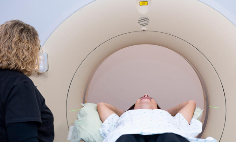
Its tests permit your primary care physician to look at your bones and inward organs in a harmless manner. Tests utilized in diagnosing cancer might incorporate a mechanized tomography (CT) check, bone output, attractive reverberation imaging (X-ray), positron outflow tomography (PET) sweep, ultrasound and X-beam, among others.
Biopsy.
During a biopsy, your primary care physician gathers an example of cells for testing in the lab. There are multiple approaches to gathering an example. Which biopsy system is ideal for you relies upon your sort of cancer and its area. Much of the time, a biopsy is the best way to conclusively analyze cancer.
In the research facility, specialists see cell tests under the magnifying lens. Typical cells look consistent, with comparative sizes and systematic association. Cancer cells look less systematic, with shifting sizes and without evident association.
Cancer stages
Whenever cancer is analyzed, your primary care physician will attempt to decide the degree (phase) of your cancer. Your primary care physician utilizes your cancer’s stage to decide your therapy choices and your opportunities for a fix.
Arranging tests and strategies might incorporate imaging tests, like bone outputs or X-beams, to check whether cancer has spread to different pieces of the body.
Bigger numbers show a further developed cancer. For certain sorts of cancer, the cancer stage is shown utilizing letters or words.
What are the various sorts of indicative imaging?
Imaging may likewise be utilized while performing biopsies and other surgeries like Penis enlargement in Dubai. There are three kinds of imaging utilized for diagnosing cancer: transmission imaging, reflection imaging, and emanation imaging. Each utilizations an alternate interaction.
Transmission imaging
The pillar goes rapidly through less thick kinds of tissue like watery discharges, blood, and fat, leaving an obscured region on the X-beam film. Muscle and connective tissues (tendons, ligaments, and ligaments) seem dark. Bones will seem white.
- X-beam
Figured tomography check (likewise called a CT examination or registered pivotal tomography or Feline sweep)
Bone output
Lymphangiogram (Slack)
Mammogram
Reflection imaging
These sound waves “bob” off of the different sorts of body tissues and designs at different paces, contingent upon the thickness of the tissues present.
Discharge imaging
Atomic medication utilizes the outflow of atomic particles from atomic substances brought into the body explicitly for assessment. Attractive reverberation imaging (X-ray) utilizes radio waves with a machine that makes serious areas of strength for a field that thusly makes cells radiate their own radio frequencies.


