What Are the 4 Stages of Embryonic Development?

Embryogenesis arises to be the procedure by which the embryo structures as well as develops. In mammals, the phrase does relate chiefly to the first phases of prenatal growth, whereas the words fetus, as well as foetal growth, illustrate later phases of pregnancy.
Embryogenesis does begin with the fertilisation of the egg cell by a sperm cell. There are several embryology courses through which you can understand more about embryogenesis.
In the context of placental mammals, the acrosome consists of digestive enzymes that are utilised to begin the breakdown of the extracellular matrix wrapping the egg. Therefore enabling the cell membrane of the sperm to link with the egg.
The uniting of these two cellular membranes creates a space in which the sperm cell embryo can be transmitted into the middle of the ovum. Where the nucleus coverings of both the sperm and egg cells start to lessen. The two haploid genomes come jointly to create a unique diploid genome.
Which Arrives Early in Embryonic Growth?
The first phases of embryonic growth do start with fertilisation. The procedure of fertilisation is very tightly regulated to assure that only one sperm fuses with one egg. After fertilisation, the zygote does undergo separation to set the blastula.
-
Fertilisation
Fertilisation is the pact of the female gamete (egg) and the male gamete (spermatozoa). Whether it arises normally inside the female reproductive system or with the allowance of reproductive technologies outside of the mortal body, the product is a hierarchy called a zygote.
When a woman is ovulating she discharges one egg into her Fallopian tubes (or extra in the possibility of fraternal twins). During this period, a woman’s cervical mucus will narrow, in practice for sperm to pass through extra effectively.
Following spermatozoa ejaculation inside the vagina, unique secretions enable them to swim through the cervix towards the uterine duct where fertilization takes place within 24-72h.
-
Blastocyst Development
Shortly after fertilization, the embryo is developed from a minor group of cells that are always halving inside of a detailed pattern named the blastocyst. It is shaped by two groups of cells, interior and exterior cells, and liquids.
The blastocyst keeps up inside a defensive mask during development named zona pellucida, which could be characterised as an eggshell. The external cells are found right below this mask. Which will establish the future placenta and encircling tissues to assist fetal growth in the uterus.
The internal cells of the blastocyst will become the unique tissues and parts of the human body, such as bones, muscles, membranes, the liver, and the heart.
-
Blastocyst Implantation
When the blastocyst attains the uterus it injects into the endometrium, the mucus membrane which strands the uterus. The outer cells of the blastocyst and the uterine interior lining, jointly, will establish the future placenta. The placenta is a pattern that transfers nutrients to the baby and extracts his/her excretes.
-
Embryo Development
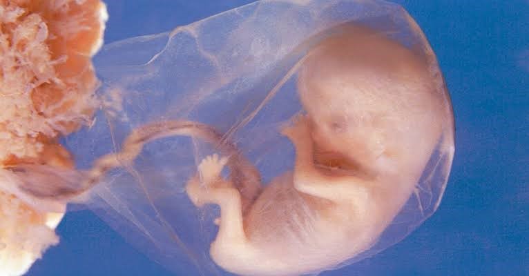
As the blastocyst enters the last phases in the implantation procedure into the internal lining of the uterus, it develops into a pattern named an embryo. This is the moment when inner organs and outer hierarchies assemble.
The mouth, lower jaw, and throat are arising, while the blood circulation procedure begins its development and a heart tube is developed. The ears originate and arms, legs, fingers, toes, and eyes are formed.
The brain and the spinal cord are already created, while the digestive region and receptive parts start their growth. The major bones are rebuilding the cartilage. Many embryology courses in India will help you to deal with the problems.


